Upper Back Anatomy ~ Upper Back Pain Center Symptoms Causes Treatments. The cervical spine protects the nerves connecting to. The superficial and intermediate muscles do not develop in the back, and are classified as extrinsic muscles. The range of motion in the upper back is limited because of the spine's attachments to the ribs (rib cage). The upper back muscles of the rhomboids and the trapezius are responsible for many of the movements of the scapula which in turn plays a huge role in the stability and mobility of the shoulder. It is like that for several reasons, all of which you can understand by looking at the anatomy of the thoracic spine.
The cause may be poor posture (such as forward head posture) or any type of irritation of the large back and shoulder muscles, including muscle strain or spasms. Anatomy of the upper back muscles. Before giving our recommendations for upper back exercises, it's important to first go over the anatomy of the back musculature. The upper back is the region below the cervical spine (neck) and above the low back (lumbar spine). The back is found posteriorly and includes the vertebral column, the muscles that support the back and the spinal cord.
Anatomy Of The Back And Spine 101 Pain Management Clinics In Las Vegas Nevada Comprehensive Pain Center from nvcpc.com This is my video about the muscles of the back. As the spine is what determines the shape of the back, as well as the structure that gives rise to a large number of the back's muscles, it is perhaps the most important component of the anatomy of the back. There is a set of muscles in the upper back (called the thoracic area) called the spinalis thoracis. The back is the body region between the neck and the gluteal regions. Extending from the base of the skull into the pelvis, it consists of 33 stacked bones known as vertebrae. License image the deltoid, teres major, teres minor, infraspinatus, supraspinatus (not shown) and subscapularis muscles (not shown) all extend from the scapula to the humerus and act on the shoulder joint. The bones of the chest and upper back combine to form the strong, protective rib cage around the vital thoracic organs such as the heart and lungs. Think of your spine as a tree trunk.
Related posts of upper back muscle diagram skeletal muscle anatomy youtube.
The bones of the chest and upper back combine to form the strong, protective rib cage around the vital thoracic organs such as the heart and lungs. Related posts of upper back muscle diagram skeletal muscle anatomy youtube. The lumbar region of the spine, more commonly known as the lower back, is situated between the thoracic, or chest, region of the spine, and the sacrum. As a result of overuse or strenuous activity, at times these tendons tend to get inflamed resulting in painful symptoms. The upper back is the region below the cervical spine (neck) and above the low back (lumbar spine). The vertebral column consists of 33 vertebrae which can be split up into 5 continuous sections. Skeletal muscle anatomy youtube 12 photos of the skeletal muscle anatomy youtube skeletal muscle anatomy youtube, human muscles, skeletal muscle anatomy youtube This muscle is located on the upper portion of the back anatomy, underneath the trapezius. Back muscles anatomy here include the trapezius, latissimus dorsi, rhomboid and levator scapulae. The cause may be poor posture (such as forward head posture) or any type of irritation of the large back and shoulder muscles, including muscle strain or spasms. Before giving our recommendations for upper back exercises, it's important to first go over the anatomy of the back musculature. The superficial and intermediate muscles do not develop in the back, and are classified as extrinsic muscles. Think of your spine as a tree trunk.
The seventh cervical vertebra, referred to as c7, meets the first of 12 thoracic vertebrae t1 at the base of the neck, a. They originate from the vertebrae and insert into the scapulae. As the spine is what determines the shape of the back, as well as the structure that gives rise to a large number of the back's muscles, it is perhaps the most important component of the anatomy of the back. The rib cage also anchors the bones of the head, neck, shoulders, and arms to the trunk of the body. The main superficial muscles of the back are the following:
8 Tips To Improve Upper Back Mass Fitness And Power from www.fitnessandpower.com The superficial and intermediate muscles do not develop in the back, and are classified as extrinsic muscles. The lumbar region of the spine, more commonly known as the lower back, is situated between the thoracic, or chest, region of the spine, and the sacrum. Related posts of upper back muscle diagram skeletal muscle anatomy youtube. The back functions are many, such as to house and protect the spinal cord, hold the body and head upright, and adjust the movements of the upper and lower limbs. It runs from the neck to the upper back. It is very stiff, and the thoracic spine has a limited range of motion. The cervical spine is the top part of the spine. Both the deltoid and the trapezius are firmly attached to …
Each block is separated by a disc that sits in between and each vertebra has a facet joint on either side.
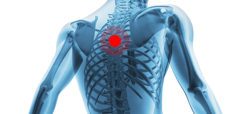
Upper back pain is most commonly caused by muscle irritation or tension, also called myofascial pain. The upper back muscles of the rhomboids and the trapezius are responsible for many of the movements of the scapula which in turn plays a huge role in the stability and mobility of the shoulder. In the upper back region, the trapezius, rhomboid major, and levator scapulae muscles anchor the scapula and clavicle to the spines of several vertebrae and the occipital bone of the skull. Powerful muscles that move the head and arms attach to these bones as well. The lumbar region of the spine, more commonly known as the lower back, is situated between the thoracic, or chest, region of the spine, and the sacrum. It consists of seven vertebrae. Upper back pain is most commonly caused by muscle irritation or tension, also called myofascial pain. The cervical spine is the top part of the spine. The main superficial muscles of the back are the following: Both the deltoid and the trapezius are firmly attached to … Skeletal muscle anatomy youtube 12 photos of the skeletal muscle anatomy youtube skeletal muscle anatomy youtube, human muscles, skeletal muscle anatomy youtube In the upper back region, the trapezius, rhomboid major, and levator scapulae muscles anchor the scapula and clavicle to the spines of several vertebrae and the occipital bone of the skull. Anatomy of the upper back muscles.
Extending from the base of the skull into the pelvis, it consists of 33 stacked bones known as vertebrae. The vertebral column consists of 33 vertebrae which can be split up into 5 continuous sections. The traps) the latissimus dorsi (a.k.a. The rib cage also anchors the bones of the head, neck, shoulders, and arms to the trunk of the body. The upper back has the most structural support, with the ribs attached firmly to each level of the thoracic spine and very limited movement.
Trapezius Muscle Stiff Neck Headache Eye Jaw Pain The Wellness Digest from thewellnessdigest.com As a result of overuse or strenuous activity, at times these tendons tend to get inflamed resulting in painful symptoms. The rhomboid muscle is activated as you bring and squeeze your scapula or shoulder blades back and together. The upper back is called the thoracic spine, and it is the most stable part of the spine. Skeletal muscle anatomy youtube 12 photos of the skeletal muscle anatomy youtube skeletal muscle anatomy youtube, human muscles, skeletal muscle anatomy youtube The back is the body region between the neck and the gluteal regions. The trapezius and latissimus dorsi muscles connect the upper limb to the vertebral column. The vertebral column consists of 33 vertebrae which can be split up into 5 continuous sections. The upper back is the region below the cervical spine (neck) and above the low back (lumbar spine).
The traps) the latissimus dorsi (a.k.a.
It runs from the neck to the upper back. The upper back is called the thoracic spine, and it is the most stable part of the spine. The rhomboid muscle is activated as you bring and squeeze your scapula or shoulder blades back and together. The vertebral column consists of 33 vertebrae which can be split up into 5 continuous sections. It consists of seven vertebrae.it comprises the vertebral column (spine) and two compartments of back muscles; The iliocostalis muscles are furthest from the spine. The cervical spine supports the weight and movement of your head and protects the nerves exiting your brain. Both the deltoid and the trapezius are firmly attached to … Upper back pain is most commonly caused by muscle irritation or tension, also called myofascial pain. The cervical spine protects the nerves connecting to. There is a set of muscles in the upper back (called the thoracic area) called the spinalis thoracis. The muscles of the back. The lower back (lumbar vertebrae) allows for flexibility and movement in back bending (extension) and forward bending (flexion).
Share :
Post a Comment
for "Upper Back Anatomy ~ Upper Back Pain Center Symptoms Causes Treatments"


Post a Comment for "Upper Back Anatomy ~ Upper Back Pain Center Symptoms Causes Treatments"